
A largely overlooked structure inside our cells may play a crucial role in how the brain forms, offering new insight into developmental disorders and potential therapies.
In a study published in Cell Reports, biomedical scientist Xuecai Ge at the University of California, Riverside and her team focused on the primary cilium, a microscopic, antenna-like structure found in nearly every cell in the human body. Despite its ubiquity, the primary cilium has remained surprisingly understudied.

“Even many biologists aren’t familiar with it,” said Ge, an associate professor of biomedical sciences in the School of Medicine. “We still have a lot to learn about this organelle.”
For decades, scientists believed the primary cilium was an evolutionary leftover with little function. But mounting evidence suggests otherwise. When the structure is disrupted, it can lead to a group of conditions known as ciliopathies, which affect multiple organs, including the brain.
“Patients may have kidney problems or obesity,” Ge said, “but when you look at their brain structure, you often see abnormalities. That made us wonder if the cilium had a role to play in brain development.”
To investigate, Ge’s team examined neural progenitor cells — early-stage cells that give rise to neurons. Each of these cells contains a single primary cilium that extends into the ventricles, fluid-filled cavities in the developing brain.
Using a large-scale biochemical approach, the researchers analyzed more than 1,000 mouse embryonic brains to identify which proteins are present in these cilia. What they found challenged existing assumptions.
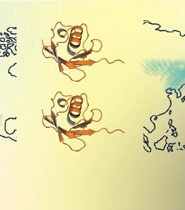
“We discovered many proteins that no one expected to find in the cilium,” Ge said. “And in some cases, these proteins are directly linked to human developmental disorders.”
One such protein, CKAP2L, is associated with Filippi syndrome, a condition that leads to reduced brain size. When the researchers removed this protein in mice, the animals developed smaller brains.
The team also found that cilia differ depending on where they are in the brain.
“We identified over 40 proteins that vary between brain regions,” Ge said. “That suggests the cilium has specialized roles, not just a single uniform function.”
For Ge, the most surprising discovery was evidence that protein production might occur directly inside the cilium itself — a concept that challenges long-standing scientific beliefs.
“The field has assumed that all proteins are made elsewhere in the cell and then transported into the cilium,” Ge said. “But we found the machinery that could make proteins on-site. It’s like finding a bread maker where you thought bread could only be delivered.”
While further research is needed to confirm whether this machinery is active, the finding could represent a major shift in how scientists understand cellular function.
The implications extend beyond basic biology, according to Ge. Because ciliopathies can affect vision, organ function, and brain development, the research could help explain how these diseases arise and how they might be treated, she said.
“Understanding which proteins are in the cilium and what they do gives us a roadmap,” Ge said. “It helps us connect genetic mutations to the actual biological processes that go wrong.”
Looking ahead, Ge’s team plans to investigate which proteins are produced within the cilium.
“We’ve only scratched the surface,” she said. “There’s a lot more to learn about how this tiny structure shapes the developing brain.”
Ge was joined in the research by scientists at UC Merced and the Scripps Research Institute in La Jolla, California.
The study was supported by grants from the National Institutes of Health and National Science Foundation.
The title of the paper is “Proximity labeling proteomics maps radial glial ciliary proteins across the developing telencephalon.”
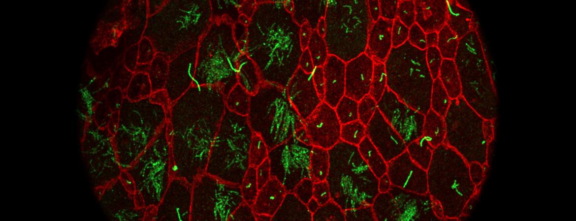
Header image shows primary cilia in the mouse embryonic brain. Cilia are in green; the cells’ boundaries are in red. (UCR/Ge lab)