
For the first time, scientists have grown functional, brain-like tissue without using any animal-derived materials or added biological coatings. The development opens the door to more controlled and humane neurological drug testing.

Neural tissue engineering’s overall goal is to create something that closely resembles the structure and function of the human brain, enabling more reproducible neurological disease studies and drug testing.
“One of the drawbacks of most brain tissue platforms is that they utilize biological coatings to help living cells thrive. These animal-derived coatings are poorly defined, which makes it difficult to recreate their exact composition for reliable testing,” said Iman Noshadi, a UCR associate professor of bioengineering who led the team.
In addition, using animal brains to conduct research relevant to human conditions — as is currently the norm — is not ideal. There are significant genetic and physiological differences between rodent and human brains. This platform could reduce, and in some cases eliminate, the need to use animal brains for this purpose and aligns with U.S. FDA efforts to phase out animal testing requirements in drug development.
The new material, described in the Advanced Functional Materials journal, functions as a scaffold on which to grow donor brain cells and could be used to model traumatic brain injuries, strokes, or neurological diseases like Alzheimer’s.
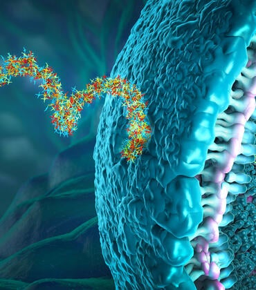
It is primarily composed of a common polymer known for its chemical neutrality called polyethylene glycol, or PEG. Typically, living cells do not attach to PEG without the addition of proteins like laminin or fibrin.
By reshaping PEG into a maze of textured, interconnected pores, the research team turned an inert material into a matrix that cells recognize, colonize, and use to build functional neural networks. Once these cells mature, they could exhibit donor-specific neural activity, allowing direct evaluation of drugs targeted to their neurological conditions.
“Since the engineered scaffold is stable, it permits longer-term studies,” said Prince David Okoro, the study’s lead author and a doctoral candidate in Noshadi’s lab. “That’s especially important as mature brain cells are more reflective of real tissue function when investigating relevant diseases or traumas.”
To build the scaffold structure, the team used a process involving water, ethanol, and PEG flowing through nested glass capillaries. When the mixture reached an outer water stream, its components began to separate. A flash of light stabilized this separation, locking in the porous structure.
The pores allow oxygen and nutrients to circulate throughout the structure efficiently, essentially feeding the donated stem cells.
“The material ensures cells get what they need to grow, organize, and communicate with each other in brain-like clusters,” Noshadi said. “Because the structure more closely mimics biology, we can start to design tissue models with much finer control over how cells behave.”
The research began in 2020 and was supported by Noshadi’s startup funds from UC Riverside. Okoro’s work was funded by the California Institute for Regenerative Medicine.
Currently, the scaffold material is only about two millimeters wide. Going forward, the team is working to scale the model and has submitted a related paper focused on liver tissue.
The group’s long-term goal is to develop a suite of interconnected organ-level cultures that reflect how systems in the body interact. They hope these tissue platforms will offer stability, longevity, and functionality comparable to the brain tissue model.
“An interconnected system would let us see how different tissues respond to the same treatment and how a problem in one organ may influence another. It is a step toward understanding human biology and disease in a more integrated way,” Noshadi said.